The neck is one of the most flexible organs of the body that has a great deal of functionality. It is quite susceptible to injuries, because it is subjected to a lot of stress and weight and to a certain extent because of its flexibility. Neck pain is usually caused by the stress, misuse, overuse, poor posture, accident or a sport injury. Highly contracted muscles and poor blood flow to the neck are the main reasons of various neck problems. Neck pain is one of the most common complaints that may become a constant problem if remain untreated and ignored.
In holistic medicine, massage therapy is considered as the most significant natural treatment that helps provide effective pain management. Massage therapy is the natural way of treating neck pain. It is a profound and non-invasive holistic approach that is effective for people of all age group. It not only provides pain relief and management, but also decelerates the progression of neck problems.
NECK INJURIES
Pain in the back is common. It can be acute (of sudden onset and painful) or can gradually come on through poor posture and other factors. A lot of pain that occurs in the back is due to muscles becoming too tight. The tight muscles do not allow as much blood into them as is needed and therefore they do not get the energy and nutrients they need to stay healthy. Tight muscles in the back is a chronic condition which develops gradually, slowly getting worse until the individual seeks treatment.
-
Overuse causes small micro tears in the muscles. The muscles then tighten up to protect themselves.
-
Poor stretching routines, particularly after training. If the muscle is not stretched to it’s natural length regularly it may adaptively shorten.
-
Scoliosis. If you have a sideways curve in the spine then some muscles will be put under more strain than they can cope with.
-
Bad posture. The head is a very heavy object and if you position it just a few centimetres the wrong way this can considerably increase the work the muscles of the back and neck have to do
Use sports massage for treatment and also stretching techniques as part of a rehabilitation programme. Concentrate on good posture until it becomes second nature. A good taping method can encourage you to maintain correct posture.
BACK INJURIES
SHOULDER INJURIES
The shoulder is one of the largest and most complex joints in the body. The shoulder joint is formed where the humerus (upper arm bone) fits into the scapula (shoulder blade), like a ball and socket. The shoulder has several other important structures:
-
The rotator cuff is a collection of muscles and tendons that surround the shoulder, giving it support and allowing a wide range of motion.
-
The bursa is a small sac of fluid that cushions and protects the tendons of the rotator cuff.
-
A cuff of cartilage called the labrum forms a cup for the ball-like head of the humerus to fit into.
-
The humerus fits relatively loosely into the shoulder joint. This gives the shoulder a wide range of motion, but also makes it vulnerable to injury.
Frozen shoulder, shoulder tendonitis, dislocated shoulder and a broken collar bone are extremely common shoulder injuries. Shoulder injury symptoms typically include shoulder pain, shoulder weakness, a stiff shoulder and shoulder joint instability. Shoulder pain and shoulder injuries frequently occur due to trauma or sporting overuse.
ELBOW INJURIES
Tennis elbow
Pain on the outside of the elbow when extending the wrist and fingers.
Golfers elbow
Pain on the inside of the elbow when flexing the wrist and fingers.
Dislocated elbow
A separation of the bones at the elbow caused by a traumatic injury and usually involving factures.
Ulna plate fracture
A fracture of the growth plate of the ulna bone, close to the elbow joint.
Biceps tendinitis at elbow
An overuse injury of the thick biceps tendon at the front of the elbow.
Radial nerve entrapment
An injury with similar symptoms to tennis elbow, other than tingling or numbness in the outer forearm or hand.
Bruised elbow
leeding in the joint caused by soft tissue damage from an impact.
There are some other forms of Elbow Injuries.
Here are some named below.
Students elbow
Also known as olecranon bursitis, a swelling develops over the elbow bone.
Medial collateral ligament sprain
njury to the medial ligament of the elbow causes pain on the inner joint, usually after a forceful injury.
Elbow hyperextension injury
An injury caused by over-straightening the elbow.
Ulna nerve injury
The ulnar nerve runs down the inner siede of the elbow and causes the ‘funny bone’ pain when knocked.
Osteochondritis dessicans in elbow
A fragmentation of the cartilage within a joint causing loose bodies and possible locking of the joint.
Triceps tendon insertion inflammation
An overuse injury causing pain at the back of the elbow, especially on resisted elbow extension.
Use manual therapy treatments such as massage therapy, myofacial release and/or transverse friction techniques across the tendon.
WRIST INJURIES
The wrist is a wonderful tool and works with your hand, forearm and elbow. You can circle your wrist and it will move forewords and backwards and this movement is necessary in many racquet sports, especially the cocking of the wrist in badminton. It is therefore important to learn correct technique to avoid injury. In volleyball the wrist is involved in powerful movements, and in canoeing a repetitive action, also golf wrist injuries are common. It is no wonder that the wrist can be not only sprained or strained but develops wrist pain from other causes.
A wrist sprain is a torn or partially torn ligament and it is possible to sprain several ligaments at one time and a strain is a torn or partially torn tendon also of which one or more can be damaged. Overuse injury usually creeps up on you and is perhaps the ligament or tendon being over stretched. Using specialised skills in assessment and treatment, the therapy will help to prevent injury or impairment, restore functional activity, and enhance participation in your daily life.
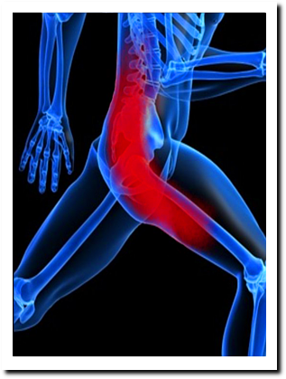
HIP INJURIES
The hip is the most stable joint in the body and is well protected. It is surrounded by muscle on all sides, but has great range of motion. This ball and socket joint lies between the head of the femur and the acetabulum of the pelvis. The hip has multiple muscle attachments (back, abdomen, hamstrings, quadriceps, abductors and adductors, and gluteal muscles). Most of the muscles of the hip are shorter and fatter than those of the leg, and allow rotation and help stabalize the joint.
Most of the hip injuries result from these small muscles being overused or pushed to hard.
Common injuries: Adductor Tendinitis or groin strain, gluteus medius tendonitis, quadriceps tendonitis, hamstring tendonitis, piriformis hypertension, sciatic nerve entrapment.
One way to encourage muscle repair is with sports massage. Sports massage will help release any built up tension and lactic acid in the overworked muscles so that blood and oxygen can return to the muscle and effectively promote muscle repair. Sports massage, if received as part of a sports program, can help an athlete prevent injury to the hip due to overuse.
KNEE INJURIES
The knee joint is the largest joint in the body, consisting of 4 bones and an extensive network of ligaments and muscles. Because of its complex structure and weight-bearing capacity, the knee is the most commonly injured joint. More severe injuries include bone bruises or damage to the cartilage or ligaments. There are two types of cartilage in the knee. One is the meniscus, a crescent-shaped disc that absorbs shock between the thigh (femur) and lower leg bones (tibia and fibula). The other is a surface-coating (or articular) cartilage. It covers the ends of the bones where they meet, allowing them to glide against one another. The four major ligaments that support the knee are the anterior cruciate ligament (ACL), the posterior cruciate ligament (PCL), the medial collateral ligament (MCL), and the lateral collateral ligament (LCL). Knee injuries can result from a blow to or twist of the knee; from improper landing after a jump; or from running too hard, too much, or without proper warmup.
Knee Anatomy
The knee joint is the largest joint in the body, consisting of 4 bones and an extensive network of ligaments and muscles. Injuries to the knee joint are amongst the most common in sporting activities and understanding the anatomy of the joint is fundamental in understanding any subsequent pathology. Bones of the knee joint
The knee is made up of four main bones- the femur (thigh bone), the tibia (shin bone), fibula (outer shin bone) and patella (kneecap). The main movements of the knee joint occur between the femur, patella and tibia. Each are covered in articular cartilage which is an extremely hard, smooth substance designed to decrease the frictional forces as movement occurs between the bones. The patella lies in an indentation at the lower end of the femur known as the intercondylar groove. At the outer surface of the tibia lies the fibula, a long thin bone that travels right down to the ankle joint. The knee joint capsule
The joint capsule is a thick ligamentous structure that surrounds the entire knee. Inside this capsule is a specialized membrane known as the synovial membrane which provides nourishment to all the surrounding structures. Other structures include the infrapatellar fat pad and bursa which function as cushions to exterior forces on the knee. The capsule itself is strengthened by the surrounding ligaments. Ligaments of the knee joint
The stability of the knee owes greatly to the presence of its ligaments. Each has a particular function in helping to maintain optimal knee stability in a variety of different positions.
Medial Collateral Ligament (MCL) – This band runs between the inner surfaces of the femur and the tibia. It resists forces acting from the outer surface of the knee-valgus forces.
Lateral Collateral Ligament (LCL) – This ligament travels from the outer surface of the femur to the head of the fibula. It resists impacts from the inner surface of the knee-varus forces.
Anterior Cruciate Ligament (ACL) – The ACL is one of the most important structures in the knee- not least because injury to it may require extensive surgery and rehabilitation. The cruciate ligaments are so called because they form a cross in the middle of the knee joint. The ACL, travels from the anterior (front) of the tibia to the posterior (back) of the femur and prevents the tibia moving forward. It is most commonly injured in twisting movements.
Posterior Cruciate Ligament (PCL) – This ligament travels from the posterior surface of the tibia to the anterior surface of the femur and in doing so wraps around the ACL.
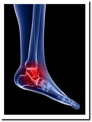
ANKLE INJURIES
Ankle sprains are some of the most common sports injuries. A sprained ankle or twisted ankle as it is sometimes known, is a common cause of ankle pain. A sprain is stretching and or tearing of ligaments (you sprain a ligament and strain a muscle). The most common is an inversion sprain (or lateral ligament sprain) where the ankle turns over so the sole of the foot faces inwards, damaging the ligaments on the outside of the ankle.
The most common damage sustained in a sprained ankle is to the anterior talofibular ligament. This ligament, as the name suggests, connects the talus (ankle bone) with the fibula (smaller of the two bones in the lower leg).
If the sprain is severe there might also be damage to the calcaneofibular ligament (connects the heel bone to the fibula) which is further back towards the heel. This ligament only becomes injured in more severe injuries due to its increased strength and laxity whilst the toes are pointed (a common position for ankle sprains).
Sports massage can be effective in treating a sprained ankle in a number of ways. Initially, light massage around the ankle, calf and shin muscles can be used to help reduce swelling from around 3 days after injury. As pain subsides, deeper techniques can be incorporated to help loosen the calf and shin muscles and improve range of motion.